

BIOLOGY - HEART STRUCTURE
Heart Anatomy Quiz
- Focuses on identifying heart valves in diagrams and images
- Covers the anatomy of the heart's chambers and valves
- Emphasizes understanding of blood flow through the heart
- Designed for Grade 9 students with a challenging difficulty level
Here are the first 3 questions. Click “View all questions” to see the complete assessment.
Q1.
Q2.
In a labelled heart schematic, L lies between the left atrium and left ventricle, M lies where blood leaves the left ventricle, N lies between the right atrium and right ventricle, and P lies where blood leaves the right ventricle. Which labels identify the semilunar valves?
Q3.
A heart diagram uses arrow A for the valve at the entrance to the artery that forms the aortic arch. Arrow B points to the valve between the right atrium and right ventricle. Which identification is correct?
Q4.
A heart image has been rotated, so page position cannot be used to identify the sides. Label R points to the valve between the chamber receiving pulmonary veins and the ventricle with the thickest muscular wall. Which valve is R?
Q5.
A flow diagram through the right side of a mammalian heart shows: vena cava → right atrium → valve T → right ventricle → valve U → pulmonary artery. How should T and U be identified?
Q6.
On a transparent heart overlay, label K points to pocket-shaped cusps with no chordae tendineae. They are located at the base of the vessel arising from the left ventricle and supplying the systemic circulation. What does K identify?
Q7.
In a sectional heart drawing, Y points to cusps attached to chordae tendineae between an atrium and a ventricle. Z points to pocket-shaped cusps without chordae at the opening from a ventricle into an artery. Which classification is correct?
Q8.
Examine the labelled longitudinal section of the mammalian heart. Which pair of labels marks the atrioventricular valves?
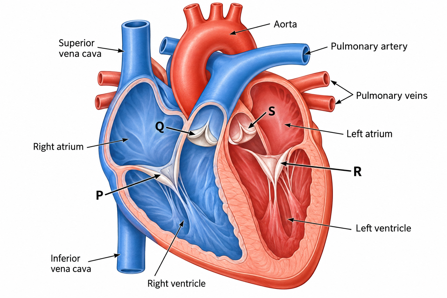
Q9.
The image shows a superior view of four valve openings after the atria and major vessels have been cut away. Which label identifies the pulmonary semilunar valve?
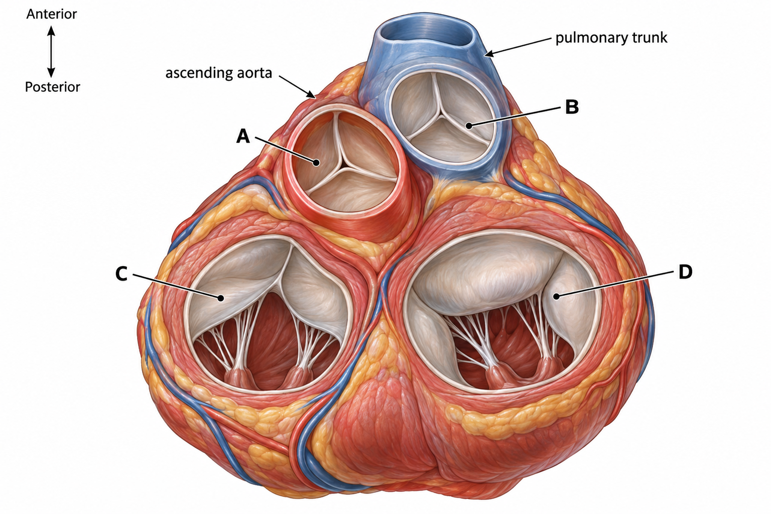
Q10.
Study the labelled photograph-style image of a dissected mammalian heart. Which label identifies the bicuspid (mitral) valve?
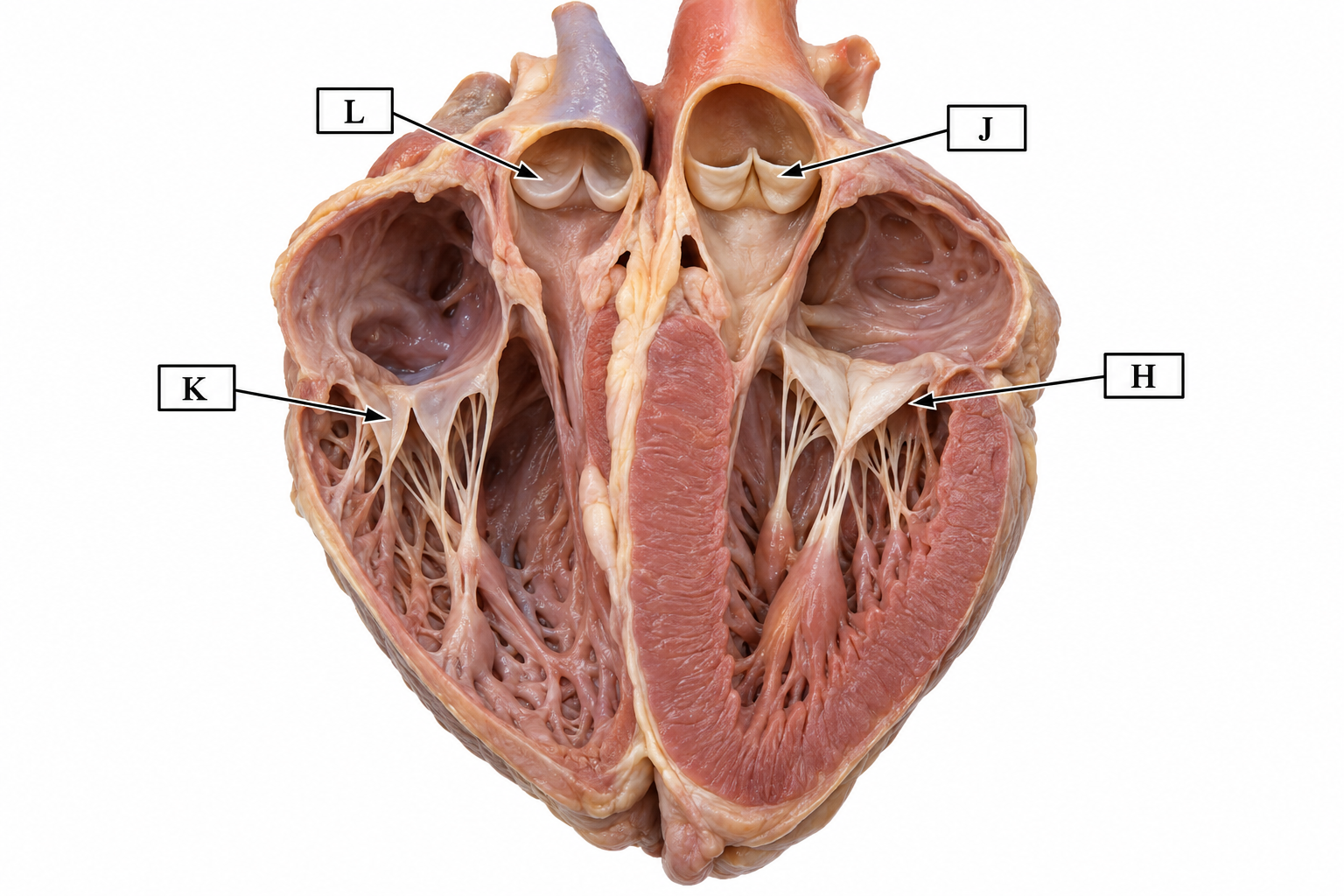
What this assessment covers
Concepts tested
- Anatomy of heart valves
- Function and location of atrioventricular valves
- Identification of semilunar valves
- Blood flow through the heart
- Structure of the heart's chambers
Question Mix
- Hard questions included
Skills you build
- Identify heart valves in diagrams
- Interpret anatomical heart images
- Differentiate between valve types
- Understand blood flow pathways
- Analyze heart structure in sectional views
Related assessments
More practice
Other assessments in this subject
